Brief Summary
This video provides a comprehensive guide on how to prepare and examine vaginal wet preps under a microscope to diagnose common vaginal infections. It covers the necessary equipment, preparation techniques for saline and KOH preps, identification of key cellular structures and microorganisms, and interpretation of test results for diagnosing trichomoniasis, bacterial vaginosis, and yeast infections.
- Explains how to prepare saline and KOH wet mounts.
- Details the identification of squamous epithelial cells, PMNs, trichomonads, clue cells, and yeast.
- Demonstrates how to use a microscope for effective slide examination.
- Outlines diagnostic criteria for common vaginal infections based on microscopic findings, whiff test, and pH levels.
Introduction
The video introduces the process of diagnosing vaginal infections through microscopic examination of vaginal samples. Clinicians check vaginal pH, examine discharge, and collect samples for further analysis under a microscope to identify the cause of symptoms like itching or unusual odour. The video aims to guide viewers on preparing and examining vaginal wet preps and performing a whiff test, which, combined with pH testing, aids in accurate diagnosis.
Equipment and Preparation
The video details the essential components of a compound light microscope, including the 10x and 40x objective lenses, the stage, condenser, light source, coarse and fine adjustment knobs, and oculars. It then explains how to prepare wet mount slides, including both a saline and a potassium hydroxide (KOH) prep. The process involves placing a drop of the vaginal sample suspension (mixed with saline) on a slide, covering it with a coverslip, and then preparing a second slide with a drop of the sample and KOH for the whiff test, which checks for a fishy or amine odour. The importance of speed is emphasised to observe trichomonads before they lose motility.
Identifying Key Cells and Organisms
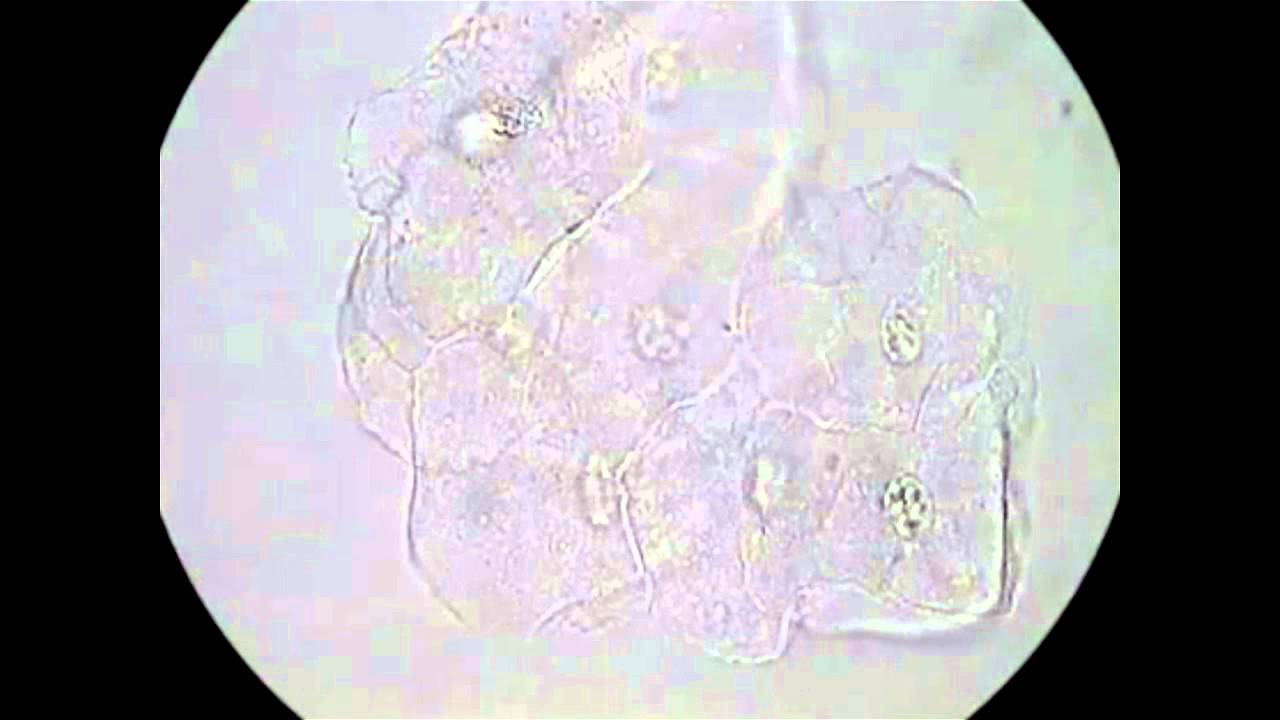
The video describes the key cells and organisms that may be found in vaginal wet mounts. Normal squamous epithelial cells are large, flat cells with a small nucleus. Polymorphonuclear leukocytes (PMNs), or white blood cells, are small, round cells with multiple nucleus lobes, indicating possible infection if found in large numbers. Trichomonads are pear-shaped protozoa identified by their jerking movement via flagella. Clue cells are squamous epithelial cells coated with bacteria, obscuring at least 75% of their borders, and are associated with bacterial vaginosis. Yeast can appear as pseudohyphae (branching, tubular forms) or budding yeast (paired cells resembling a shoe print).
Saline and KOH Preps
The video explains the utility of saline and KOH preps in identifying different microorganisms. Saline preps allow the visualisation of epithelial cells, PMNs, trichomonads, clue cells, red blood cells, sperm, and bacteria. While yeast can be seen in saline, it's sometimes obscured by other cells. KOH preps are specifically used to detect yeast by lysing epithelial cells, PMNs, and clue cells, making yeast more visible. A positive whiff test, indicated by a fishy odour, may suggest trichomoniasis or bacterial vaginosis due to the volatilisation of amines by KOH.
Microscope Usage
The video provides a step-by-step guide on using a microscope to examine wet mount slides. It includes placing the slide on the stage, using the 10x objective to initially focus with the coarse adjustment knob, adjusting the condenser or diaphragm for optimal contrast, switching to the 40x objective for higher magnification using the fine adjustment knob, and increasing illumination as needed. The importance of cleaning the objective lenses with lens cleaning solution and paper is stressed. It's recommended to spend at least three minutes reading each slide, using a Z pattern to ensure thorough examination.
Examining Patient Samples
The video demonstrates the examination of various patient samples under the microscope. It shows normal squamous epithelial cells, red blood cells, PMNs, and bacteria (likely lactobacilli). Examples of trichomonads moving in a jerky motion, clue cells with bacteria-covered surfaces, and budding yeast are presented. The video also highlights the importance of distinguishing between true clue cells and epithelial cells with some bacteria, as well as recognising artifacts. Both saline and KOH preps are used to identify pseudohyphae and budding yeast, with the KOH prep providing a clearer view due to lysed background cells.
Diagnostic Criteria
The video outlines the diagnostic criteria for common vaginal infections based on test results. For trichomonas vaginitis, the saline prep shows motile trichomonads, the KOH prep does not, the whiff test may reveal a fishy odour, and the pH is typically greater than 4.5. Bacterial vaginosis is indicated by clue cells in the saline prep (at least one in ten fields or one in five epithelial cells), a negative KOH prep for clue cells, a fishy odour in the whiff test, and a pH greater than 4.5. Yeast vaginitis is characterised by budding yeast and/or pseudohyphae in both saline and KOH preps, no fishy odour in the whiff test, and a pH generally less than 4.5. The video notes that patients may have multiple infections, requiring identification of combined microorganisms and cells for appropriate treatment.