Brief Summary
This video continues a series on the skeletal system, focusing on the nutrient foramen, blood supply to bones, types of epiphysis, and an introduction to bone tissue. It explains the importance of the nutrient foramen in bone health and surgical considerations, details the arterial blood supply to bones and its clinical implications, describes the four types of epiphysis, and provides a foundational overview of bone tissue composition, including its organic and inorganic components.
- Nutrient foramen and artery are crucial for bone nourishment and surgical safety.
- Understanding blood supply helps explain conditions like osteomyelitis.
- Epiphysis types include pressure, traction, atavistic, and aberrant forms.
- Bone tissue comprises cells and an extracellular matrix with organic and inorganic components.
Introduction and Dedication
The video begins with a warm welcome to "Real in Partition Moments" and acknowledges that this is part three of a ten-part series on the skeletal system. The presenter dedicates this episode to his father, Mr. Daniel, whom he describes as a great teacher and the "brain behind the camera." He also expresses gratitude to other influential teachers in his life, including Mr. Raymond Attubani and Mr. Artificial.
Acknowledgements
The presenter acknowledges viewers who have shown enthusiasm for the content through their comments. He mentions three individuals by name: Caleb, a first-year medicine student at KNUST; Stephanie Edu; and Nelson, a first-year student at Christian Service University. He appreciates their encouraging feedback and expresses his intention to acknowledge supportive viewers in future videos.
Nutrient Foramen and Artery
The discussion centres on the nutrient foramen and its importance. The nutrient artery supplies the medullary cavity and the inner two-thirds of the cortical bone. The nutrient foramen takes an oblique course, either downwards towards the distal epiphysis or upwards towards the proximal end of the bone. Knowledge of the nutrient foramen is crucial to avoid iatrogenic injuries during orthopaedic surgeries and to understand the growing end of the bone. Typically, one long bone has one nutrient foramen, but some individuals may present with double nutrient foramina, especially in the tibia and fibula of males, due to their greater involvement in manual labour.
Orientation of Nutrient Foramina
In upper limb bones, nutrient foramina are generally oriented anteriorly, while in lower limb bones, they are displaced at the crucial aspect. This difference is due to the 90-degree rotation of the bones during development. In long bones of the upper limb, the nutrient foramina are directed obliquely towards the elbow joint ("To the Elbow I go"). In the lower limb, they are directed away from the knee joint ("From the Knee I Flee"). The clavicle's nutrient foramen is directed towards the lateral end, making the medial end the growing end.
Clinical Significance and Bone Growth
The presenter explains that the direction of the nutrient foramen indicates the growing end of the bone. The epiphyseal plate, made of hyaline cartilage, allows for interstitial growth. The knowledge of nutrient artery direction helps predict which epiphyseal plate will fuse earlier. Once the epiphyseal plate fuses, it becomes the epiphyseal line, indicating that synostosis has occurred. Articular cartilage is also present at joint surfaces.
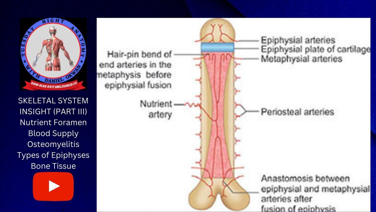
Arterial Blood Supply to the Bone
The video transitions to the arterial blood supply to the bone, emphasising the importance of understanding this for clinical reasons. The nutrient artery enters through the nutrient foramen and divides into ascending and descending branches. Epiphyseal arteries supply the epiphysis, and metaphyseal arteries supply the metaphysis. Anastomoses occur between metaphyseal arteries and nutrient arteries. In children, the epiphyseal plate prevents anastomosis, leading to hairpin bends in the metaphyseal arteries.
Osteomyelitis in Children
The lack of anastomosis in children can lead to osteomyelitis. Bacterial infections in the bloodstream can lodge in the hairpin bends of metaphyseal arteries, leading to inflammation of the bone. This explains why osteomyelitis is more common in children and occurs in the metaphyses. The presenter also touches on periosteal arteries, which supply the outer one-third of the cortical bone and are crucial for bone healing after a fracture. The periosteum has two layers: an inner cellular layer with osteogenic cells and an outer fibrous layer. Appositional growth, facilitated by the inner layer, increases the diameter of the bone.
Types of Epiphysis
The video outlines four types of epiphysis: pressure, traction, atavistic, and aberrant. Pressure epiphyses form joints, bear weight, and are covered with hyaline cartilage (e.g., head of the humerus, condyles of the femur). Traction epiphyses do not form joints but provide surfaces for muscle and ligament attachment (e.g., tubercles of the humerus, trochanters of the femur). Atavistic epiphyses are underdeveloped structures in humans that are separate bones in other quadrupeds (e.g., coracoid process of the scapula, lenticular process of incus). Aberrant epiphyses are not supposed to be present but find themselves there (e.g., secondary ossification centres in metacarpals and metatarsals).
Aberrant Epiphysis Explained
Aberrant epiphyses are further explained using miniature long bones like metacarpals, metatarsals, and phalanges. Normally, metacarpals 2-5 and metatarsals 2-5 have a primary ossification centre in the shaft and a secondary centre in the head. If a secondary centre appears in the base instead, it is considered an aberrant epiphysis. In the first metacarpal and metatarsal, the normal epiphysis is in the proximal base; if it appears in the head, it is aberrant.
Introduction to Bone Tissue
The video provides a brief introduction to bone tissue (osteous tissue), a special type of connective tissue with a mineralised extracellular matrix. Bone tissue consists of cells and an extracellular matrix, which includes inorganic and organic components. The inorganic component (mineral component) makes up about 40% of the extracellular matrix and includes hydroxyapatite. Water contributes about 25%. The organic component (protein component) makes up about 35% and includes collagenous (collagen type 1) and non-collagenous parts (osteonectin, osteopontin).
Components of Bone Tissue and Osteoporosis
The presenter details the importance of the mineral component, noting that a deficiency can lead to osteomalacia (or rickets in children). The organic component includes collagen and non-collagenous proteins. A lack of collagen can lead to osteoporosis, a condition common in post-menopausal women due to limited oestrogen production. The video concludes by previewing the next session, which will cover bone cells, types of bone tissue (spongy and compact), and ossification processes.