Brief Summary
This video by Ninja Nerd provides a comprehensive overview of cirrhosis, a condition characterized by irreversible fibrosis of the liver, leading to impaired liver function and potential portal hypertension. The video covers the pathophysiology, various causes including parenchymal, vascular, and biliary tract diseases, and the complications arising from portal hypertension and decreased liver function. It also discusses diagnostic approaches and treatment strategies, including managing complications like ascites, hepatic encephalopathy, and hepatorenal syndrome. The video emphasizes the importance of recognizing and managing these complications to improve patient outcomes and determine the need for liver transplantation.
- Cirrhosis is irreversible fibrosis of the liver, leading to decline liver function and potentially portal hypertension.
- Causes of cirrhosis include alcohol, autoimmune disorders, viral infections (hepatitis B and C), and metabolic diseases.
- Complications include portal hypertension, decreased liver function, and increased risk of hepatocellular carcinoma.
Cirrhosis Introduction
Cirrhosis is defined as the irreversible fibrosis of the liver, which leads to a decline in liver function and can result in portal hypertension. The video aims to provide a basic understanding of the pathophysiology of cirrhosis without going into excessive detail.
Pathophysiology | Parenchymal Disease
Chronic injury and damage to the liver cause hepatocytes to undergo repeated injury. This leads to the activation of stellate cells, which lay down fibrous tissue around the sinusoids, resulting in fibrosis. The liver attempts to regenerate, forming nodules, but the combination of fibrosis and nodular regeneration impairs normal liver function, leading to a decline in function, such as reduced albumin synthesis, impaired bilirubin conjugation, and decreased clearance of ammonia. The fibrous tissue also compresses portal veins, leading to portal hypertension. Common causes of direct parenchymal damage include alcohol, autoimmune hepatitis, viral infections (hepatitis B and C), and metabolic disorders like hemochromatosis, Wilson's disease, alpha-1 antitrypsin deficiency, and non-alcoholic fatty liver disease (NAFLD). Alcohol induces steatosis, while autoimmune diseases activate immune cells to destroy hepatocytes. Viral infections use liver cells to replicate, causing cell rupture and injury. Metabolic disorders lead to the accumulation of substances like iron (hemochromatosis) or copper (Wilson's disease), causing free radical damage. Alpha-1 antitrypsin deficiency results in the buildup of inactive polymers, causing direct liver injury. NAFLD causes fat accumulation, leading to inflammation and fibrosis. Liver enzymes (AST and ALT) may be elevated in early to middle stages of cirrhosis but can normalize in late stages due to the replacement of hepatocytes with fibrous tissue.
Pathophysiology | Vascular Disease
Indirect liver injury can occur due to vascular issues, such as right heart failure, which causes elevated central venous pressure and hepatic congestion, leading to chronic liver cell injury. Another cause is Budd-Chiari syndrome, characterized by clots within the hepatic veins, leading to backflow and congestion in the liver. Budd-Chiari syndrome is often associated with malignancies or genetic hypercoagulable states like Factor V Leiden or polycythemia.
Pathophysiology | Biliary Tract Disease
Chronic biliary tract diseases, such as primary biliary cirrhosis (PBC) and primary sclerosing cholangitis (PSC), can cause chronic inflammation of the intrahepatic and extrahepatic biliary ducts. This inflammation impedes bile flow, leading to bile backflow and injury to hepatocytes. PBC is characterized by intrahepatic duct inflammation only and is associated with anti-mitochondrial antibodies, while PSC involves both intrahepatic and extrahepatic duct inflammation and is often associated with ulcerative colitis.
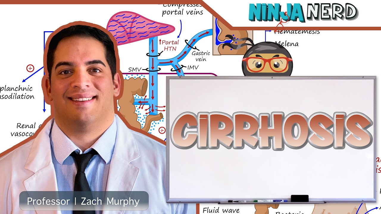
Complications | Portal Hypertension
Patients with cirrhosis can often be asymptomatic until they develop complications related to portal hypertension or declining liver function, leading to decompensated cirrhosis. Portal hypertension results from fibrous tissue compressing portal veins, causing back pressure in the superior and inferior mesenteric veins, as well as the gastric veins. This high pressure leads to the development of portosystemic shunts, where blood bypasses the liver, resulting in the buildup of ammonia in the bloodstream, causing hepatic encephalopathy. High ammonia levels lead to cerebral edema and altered mental status, characterized by confusion and asterixis. Elevated portal blood pressure in the gastric veins can cause esophageal varices, which can rupture and lead to upper GI bleeds, presenting as hematemesis or melena. Increased hydrostatic pressure in the mesenteric veins can cause fluid to leak into the peritoneal space, leading to ascites. This fluid is poor in albumin, resulting in a serum ascites albumin gradient (SAAG) greater than 1.1. Ascitic fluid can become infected, leading to spontaneous bacterial peritonitis (SBP), characterized by increasing abdominal pain, fever, and a polymorphonuclear leukocyte (PMN) count greater than 250 per millimeter cubed in the ascitic fluid. High portal blood pressure can also lead to splanchnic vasodilation, causing blood to pool in the splanchnic circulation and reducing blood flow to the systemic vascular system. This triggers the renin-angiotensin-aldosterone system and sympathetic nervous system, leading to renal artery vasoconstriction and hepatorenal syndrome, characterized by decreased urine output and increased creatinine levels.
Complications | Decreased Liver Function
When the liver fails, it can no longer synthesize albumin, leading to decreased osmotic pressure and the precipitation of ascites. The liver also produces clotting proteins (factors II, VII, IX, X) and anticoagulants (antithrombin III, protein C, protein S). Liver failure results in a greater impact on procoagulants, increasing the risk of coagulopathy and bleeding, which can manifest as intracranial hemorrhage, epistaxis, mucocutaneous bleeding, or GI bleeds. Low factor VII levels can elevate the INR. Decreased production of thrombopoietin (TPO) leads to reduced platelet production, further increasing the risk of bleeding. Liver failure also impairs ammonia clearance, leading to hepatic encephalopathy. The liver's inability to metabolize estrogen results in high estrogen levels, causing testicular atrophy, gynecomastia, palmar erythema, and spider angiomas. Impaired bilirubin conjugation and excretion lead to hyperbilirubinemia, causing jaundice.
Complications | Hepatocellular Carcinoma
Cirrhosis increases the risk of hepatocellular carcinoma due to chronic inflammation and dysplasia. Regular serial ultrasounds should be performed every six months to monitor for tumors, along with trending alpha-fetoprotein (AFP) levels, which are often elevated in patients with hepatocellular carcinoma.
Diagnostic Approach
Diagnosing cirrhosis involves labs and imaging. A complete blood count (CBC), liver function tests (LFTs), prothrombin time/international normalized ratio (PT/INR), and albumin levels should be checked. LFTs may show increased AST and ALT, though this is not always the case. Increased bilirubin, elevated PT/INR (greater than 1.5), and low albumin levels are more indicative of cirrhosis. Patients may also have low platelet counts due to decreased thrombopoietin. Imaging, such as abdominal ultrasound with elastography, can show nodular fibrosis and increased liver stiffness. A liver biopsy can definitively confirm cirrhosis and identify the underlying cause, such as autoimmune hepatitis, hemochromatosis, or Wilson's disease. To determine if a patient has decompensated cirrhosis, look for ascites, spontaneous bacterial peritonitis, esophageal varices, hepatorenal syndrome, or hepatic encephalopathy. If ascites is present, a paracentesis should be performed to check the serum albumin ascites gradient (SAAG) and neutrophil count. A SAAG greater than 1.1 suggests portal hypertension, and a neutrophil count greater than 250 indicates SBP. Patients with hematemesis or melena should be evaluated for variceal bleeding via esophagogastroduodenoscopy (EGD). Ammonia levels should be checked, though a normal level does not exclude hepatic encephalopathy. Elevated creatinine and poor urine production suggest acute kidney injury and hepatorenal syndrome.
Treatment
Treatment for cirrhosis primarily involves managing complications. For ascites, sodium restriction (less than 2 grams per day) and diuretics (spironolactone and furosemide) are used. Spironolactone, an aldosterone antagonist, blocks sodium and water retention, while furosemide removes excess sodium and water via the kidneys. For large-volume paracentesis (greater than 5 liters), albumin should be administered to prevent hypotension and hepatorenal syndrome. Refractory ascites may require a transjugular intrahepatic portosystemic shunt (TIPS), which bypasses the liver to reduce portal pressure. Spontaneous bacterial peritonitis (SBP) is treated with antibiotics, such as ceftriaxone or ciprofloxacin. Hepatic encephalopathy is managed with lactulose, which converts ammonia into ammonium for excretion, and rifaximin, which reduces ammonia-producing bacteria. Hepatorenal syndrome is treated with octreotide or midodrine, which cause splanchnic vasoconstriction to reduce renal artery vasoconstriction, and albumin to increase oncotic pressure and maintain renal perfusion. Variceal bleeding is managed with octreotide to cause splanchnic vasoconstriction, ceftriaxone to reduce mortality related to infection, and endoscopic ligation to ligate the varices. Propranolol or nadolol can be used to prevent variceal bleeds by reducing splanchnic circulation. Hepatocellular carcinoma is monitored with abdominal ultrasounds every 6 months and alpha-fetoprotein (AFP) levels. Ultimately, many patients with cirrhosis will require a liver transplant. The Child-Pugh score and MELD-Na score are used to assess the need for transplant. The Child-Pugh score assesses albumin, bilirubin, coagulation, ascites, and encephalopathy, while the MELD-Na score assesses bilirubin, INR, sodium, dialysis, and creatinine.