
Brief Summary
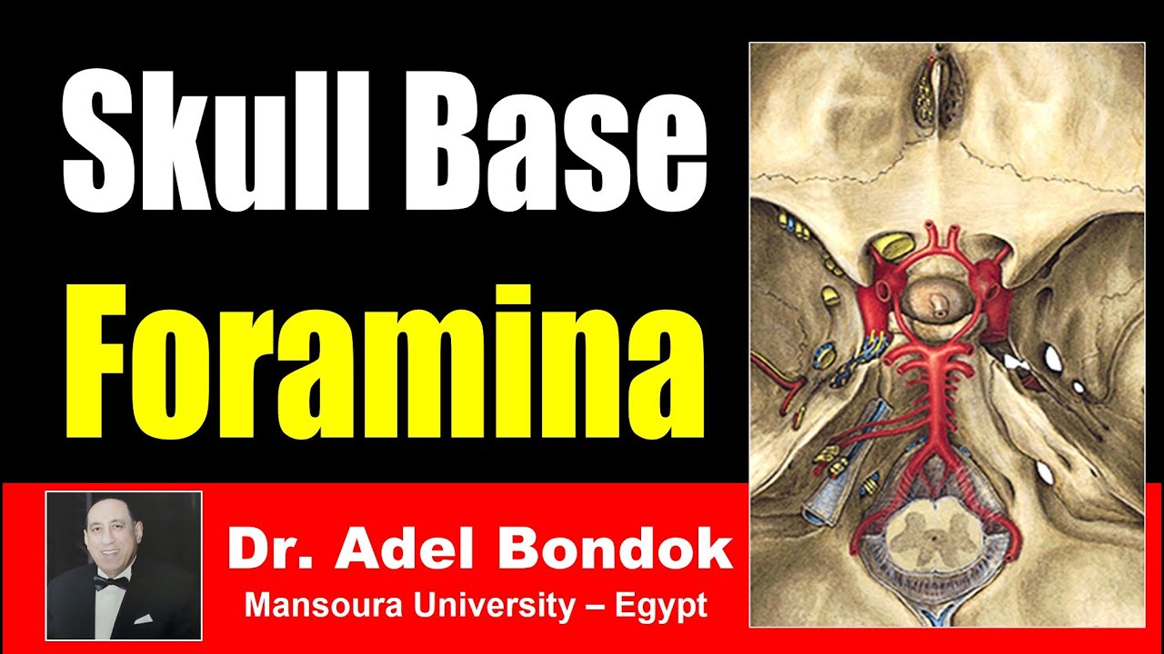
This video by Dr. Adel Bondok provides a detailed overview of the foramina (openings) of the skull and their contents, focusing on both the cranial cavity (Norma basalis interna) and the external surface of the skull base (Norma basalis externa). The lecture identifies each foramen, its location, and the specific nerves, arteries, veins, and other structures that pass through it.
- The video covers foramina in the cranial cavity, including the foramen cecum, cribriform plate, optic canal, superior orbital fissure, foramen rotundum, foramen ovale, foramen spinosum, foramen lacerum, internal auditory meatus, jugular foramen, hypoglossal canal, and foramen magnum.
- It also discusses foramina in the Norma basalis externa, such as the incisive foramen, greater and lesser palatine foramina, foramen ovale, foramen spinosum, foramen lacerum, carotid canal, jugular foramen, stylomastoid foramen, mastoid foramen, foramen magnum, hypoglossal canal, and posterior condylar canal.
Introduction
Dr. Adel Bondok, Professor of Anatomy and Neuroscience at Montsouro University in Egypt, introduces a discussion on the foramina of the skull and their contents, beginning with the foramina found within the cranial cavity.
Foramina in the Cranial Cavity (Norma Basalis Interna)
The lecture begins by identifying the foramen cecum, which transmits a vein connecting the superior sagittal sinus and the nasal veins. Next, the cribriform plate of the ethmoid bone is discussed, which transmits the olfactory nerves. The anterior and posterior ethmoidal canals, related to the cribriform plate, transmit the anterior and posterior ethmoidal nerves and vessels, respectively. The optic canal transmits the optic nerve, surrounded by three meningeal layers, and the ophthalmic artery, a branch of the internal carotid artery.
Superior Orbital Fissure
The superior orbital fissure, located between the lesser and greater wings of the sphenoid bone, transmits seven nerves and ophthalmic veins. These include the lacrimal, frontal, trochlear, superior division of the oculomotor, nasociliary, inferior division of the oculomotor, and abducent nerves, as well as ophthalmic veins.
Foramen Rotundum and Ovale
The foramen rotundum transmits the maxillary nerve, a branch of the trigeminal nerve. The foramen ovale transmits four structures: the mandibular nerve, the accessory meningeal artery, the lesser petrosal nerve, and emissary veins connecting the cavernous sinus with the pterygoid venous plexus.
Foramen Spinosum and Lacerum
The foramen spinosum transmits the middle meningeal artery and the nervus spinosus, which is the meningeal branch of the mandibular nerve. The foramen lacerum transmits the internal carotid artery after it exits the carotid canal, surrounded by a sympathetic plexus that forms the deep petrosal nerve.
Internal Auditory Meatus and Jugular Foramen
The internal auditory meatus transmits the facial nerve, vestibulocochlear nerve, and the labyrinthine artery, which supplies the inner ear. The jugular foramen is divided into three compartments, transmitting two veins, two arteries, and three nerves. The anterior compartment transmits the inferior petrosal sinus, the posterior compartment transmits the sigmoid sinus (continuing as the internal jugular vein), and the middle compartment transmits the glossopharyngeal, vagus, and accessory nerves, along with meningeal branches of the occipital and ascending pharyngeal arteries.
Hypoglossal Canal and Foramen Magnum
The hypoglossal canal transmits the hypoglossal nerve. The foramen magnum transmits at least nine structures: the medulla oblongata (becoming the spinal cord), spinal accessory nerves, sympathetic fibers around the arteries, meningeal branches of the upper three cervical nerves, vertebral arteries, anterior spinal artery, posterior spinal arteries, and the three meningeal layers (dura mater, arachnoid mater, and pia mater).
Foramina in the Norma Basalis Externa: Incisive and Palatine Foramina
The lecture transitions to the external surface of the skull base. The incisive foramen transmits the sphenopalatine artery and the nasopalatine nerve. The greater palatine foramen transmits the greater palatine nerve and vessels, while the lesser palatine foramen transmits the lesser palatine nerve and vessels.
Foramen Ovale and Spinosum (Revisited)
The foramen ovale again is noted to transmit the mandibular nerve, accessory meningeal artery, lesser petrosal nerve, and emissary veins. The foramen spinosum transmits the middle meningeal artery and the nervus spinosus.
Foramen Lacerum and Carotid Canal
The foramen lacerum transmits the internal carotid artery, surrounded by a sympathetic plexus forming the deep petrosal nerve, after the artery exits the carotid canal. The carotid canal itself transmits the internal carotid artery surrounded by a sympathetic plexus.
Stylomastoid and Jugular Foramina
The stylomastoid foramen, located between the styloid and mastoid processes, transmits the facial nerve and the stylomastoid artery, a branch of the posterior auricular artery. The jugular foramen is again described with its three compartments, transmitting the inferior petrosal sinus, sigmoid sinus, glossopharyngeal, vagus, and accessory nerves, along with meningeal branches of the ascending pharyngeal and occipital arteries.
Mastoid Foramen and Foramen Magnum (Revisited)
The mastoid foramen transmits a meningeal branch of the occipital artery and an emissary vein connecting the occipital veins and the sigmoid sinus. The foramen magnum is reiterated to transmit the medulla oblongata, spinal accessory nerves, sympathetic fibers, meningeal branches of cervical nerves, vertebral arteries, spinal arteries, and meningeal layers.
Hypoglossal and Posterior Condylar Canals
The hypoglossal canal transmits the hypoglossal nerve. The posterior condylar canal transmits an emissary vein connecting the sigmoid sinus and the suboccipital plexus of veins.